Radiographic anatomy of the thoracic limb of the Ladoum sheep
Published 2025-07-09
Keywords
- Array,
- Array,
- Array,
- Array,
- Array
- Array,
- Array ...More
Copyright (c) 2025 Moroccan Journal of Agricultural and Veterinary Sciences

This work is licensed under a Creative Commons Attribution-NonCommercial-ShareAlike 4.0 International License.
Abstract
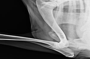
This study, which took place between January 2021 and April 2022, involved a group of 15 randomly selected Ladoum sheep. After a clinical examination at the Medina Veterinary Clinic to ensure the animals were healthy, radiographs were taken at the Bombo Clinic. This study made it possible to create a radioanatomical atlas of the thoracic limb of sheep to highlight the specific features of the shoulder joint. The humerus has a head separated from the body by a slightly marked neck and the elbow joint, where there is a slight enlargement corresponding to the synovial fossa of the ulna, the two rows of carpals, and the two fingers (phalanges).
Keywords: Radiography, Limbs, Thoracic, Ladoum Sheep, Senegal